Monday, December 2, 2019


Cirrhosis of the Liver
by Laura Kowalk Rogge RN, BSN & Carlyn Husbands RN, BSN

Cirrhosis of the liver is a disease in which the hepatic cells become damaged and scarred. The two most common causes are excessive alcohol use and viral infections of the liver such as hepatitis. Other causes can be autoimmune disorders, disorders of the bile duct and obesity, uncontrolled hyperlipidemia and diabetes. 
The liver has a few very important functions: 1) metabolizes, 2) detoxifies, 3) stores and 4) produces. An interruption is any of these hepatic functions can cause the cells in the liver to die and become fibrous, leading to irreversible liver damage called cirrhosis. In addition, this fibrous scar tissue interferes with the blood flow of the liver resulting in portal hypertension. Portal hypertension is a serious complication of cirrhosis that can lead to splenomegaly and bleeding varices within the stomach, esophagus and/or rectum which can be potentially fatal.
During metabolism the liver will break down waste products and convert them into something that the body can use. An example of this is when the body metabolizes ammonia into urea. Ammonia, a by-product of protein metabolism, will go to the liver to be metabolized and is converted into urea which is then excreted via the kidneys as urine. In cirrhosis, the liver will be unable to convert the ammonia into urea leading to dangerous levels of elevated ammonia causing toxic hepatic encephalopathy.
The liver is responsible for detoxifying all substances we ingest, such as alcohol and medications, deciding what can pass through safely into our bloodstream and throughout our body. It does this with the help of the Kupffer cells.
Glycogen is an accumulation of glucose and is stored in the liver. When the body has an excess of glucose such as from eating a heavy meal, it will store all this extra glucose as glycogen. When the body requires energy, it will tap into this storage of glycogen and convert it to glucose for the body to use as energy. In cirrhosis, both of these storage functions can be impaired causing hyperglycemia when the liver is unable to take in and store the excess glucose. Hypoglycemia will occur when the liver is unable to convert the glycogen back to glucose when the body requires it. The liver also stores the vitamins A, C, E, D, K, B12 and iron. In cirrhosis, the liver is unable to absorb these necessary vitamins.  
Albumin, bile and coagulation factors are produced in the liver. Albumin is a necessary protein in that is attracts fluids and drugs and brings them into the vascular system. It also is bound to calcium and is important for bones. Bile is the substance that transports old red blood cells (bilirubin) to the spleen, and also transports cholesterol, flushing it out of our body via the stool. Coagulation factors such as PT, PTT, INR are produced in the liver and are responsible for the clotting of our blood.
When in the early-stages, cirrhosis symptoms can often go undetected. Frequently, cirrhosis is first discovered via routine blood work. To help substantiate the Dianosis, both lab and imaging tests are done. Liver function tests include enzymes that are found in the liver ALT AST ALP and bilirubin. Coagulation tests PT, and hepatitis antibodies are also used. An ultrasound, Ct, or MRI of abdomen may also be done. 

 Treatment for cirrhosis varies according to the cause and extent of the liver damage. The goals of treatment are to slow the progression of scarring by prevention or treating symptoms and problems cause by cirrhosis.
If you have cirrhosis caused by excessive alcohol use, try to STOP drinking. Alcohol  in cirrhosis is toxic to the liver.

Weight loss. People with cirrhosis caused by nonalcoholic fatty liver disease could become healthier if they lose weight and control their blood sugar levels.
Medications may limit further damage to liver cells caused by hepatitis B or C via specific Tx.  of the viruses.

Staff, M. C. (2018, December 07). Cirrhosis. Retrieved from mayoclinic.org: https://www.mayoclinic.org/diseases-conditions/cirrhosis/symptoms-causes/syc-20351487


Edited by Shirley Comer DNP, RN, JD, CNE, ACNS-BC, APN

Irritable Bowel Syndrome by Artria Alexander RN BSN

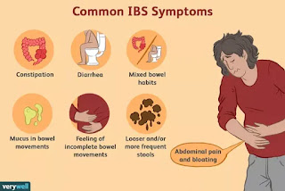
Irritable bowel Syndrome (IBS) is a common gastrointestinal disorder characterized by a cluster of signs and symptoms that effects the process of digestion in the large intestine. The process of food absorption and water absorption is compromised. IBS triad of signs and symptoms include; cramping, abdominal pain, and altered bowel habits (constipation/diarrhea). 
Signs of IBS include abdominal distention, food intolerance, and occasionally weight loss. 

Factors that contribute to IBS are linked to muscle contractions in the intestine, abnormalities in the nervous system including poor coordination between the brain and intestine, inflammation of the large intestine, and severe infections caused by bacteria or viruses. The symptoms of IBS can be triggered by food, stress and hormones. 

Those who are at an increased risk of developing IBS include; females, individuals under age 50, those with a family history of IBS, and those who have preexisting mental health problems such as anxiety and depression.

To diagnosis and treat IBS, a comprehensive health history and physical exam are done. Diagnostic criteria include; X-ray, CT scan, colonoscopy, sigmoidoscopy, lactose intolerance test, upper endoscopy, and stool test. Treatment options are geared toward lifestyle and diet modifications consisting of high fiber foods, the intake of plenty of fluids, rest and adequate sleep, and exercise. Other options for treatment include medication management such as fiber supplements (Metamucil), laxatives (Miralax), anti-diarrheal medication (Imodium), anticholinergic medications, (Bentyl), tricyclic antidepressants and pain medications (Neurontin).

Reference:
Chang, L. (n.d.). Irritable Bowel Syndrome (IBS). Retrieved November 23, 2019, from https://www.niddk.nih.gov/health-information/digestive-diseases/irritable-bowel-syndrome.

Edited by Shirley Comer DNP, RN, JD, CNE, ACNS-BC, APN


Monday, May 6, 2019


Acute Bronchitis 

Written by Judith Ose RN, BSN, MSNc and Nwachukwu Nkechi RN, BSN, MSNc, Edited by Shirley Comer 

In general, Acute Bronchitis is a viral infection whose predominant symptom is a cough which can last up to three weeks (Hart, 2014, p. 34). Moreover, this viral infection can present like a mild cold. In addition, an individual with acute bronchitis can also present with wheezing, especially if there is an underlying disease process of asthma (Hart, 2014, p. 34).

            Antibiotics are usually not needed in the treatment of acute bronchitis unless cases of high fever, high pulse rate, and respiration are involved.

Risk factors of acute bronchitis
  • Close contact with someone who has a cold or acute bronchitis
  • Failure to get age-appropriate immunizations
  • Exposure to tobacco smoke, fumes, dust, and air pollution
Diagnostic criteria and treatments. 
History is one of the main steps in diagnosing acute bronchitis. The following symptoms may help in the diagnosis of acute bronchitis; CBC with differential, procalcitonin to rule out a bacterial infection, blood culture if bacterial is highly suspected, chest x-ray in elderly patients or if physical findings are suggestive of pneumonia.

The most debilitating complication of this disease process is the actual cough and therefore the treatment focuses primarily on suppressing the cough The treatments for acute bronchitis include Dextromethorphan 20 mg PO, q4hrs or 60 mg extended-release liquid BID for the cough, inhaled bronchodilator albuterol 2.5 mg TID/QID by nebulizer, for wheezing, and Benzonatate  200mg and Guaifenesin 600mg PO q6hrs, for the mucus production (Hart, 2014, p.36). 

References 

Hart, A. (2014). Evidence-based diagnosis and management of  acute bronchitis. The Nurse Practitioner 39(9), 35-36 doi: 10.1097/01.NPR. 0000452978.99676.2b

Kinkade, S., & Long, N. (2016). Acute bronchitis. American Family Physician94(7), 560-565. Retrieved from https://www.aafp.org/afp/2016/1001/p560.pdf

Fayyaz, J. (2018). Bronchitis essential practice, background, pathophysiology. Retrieved from: https://emedicine.medscape.com/article/297108-overview   

Knutson, D., Braun, C. (2015). American family physician. Diagnosis and management of acute bronchitis retrieved from:  https://www.aafp.org/afp/2002/05/15/p2039.html           


Gout
By Kimberly Lopez RN, BSN, MSNc 
and Mia Watkins RN, BSN, MSNc
Edited by Shirley Comer

Gout is a type of arthritis that predominately affect older males, but can affect women too.  It is more common in African Americans (5%) than whites (4%) (ncbi.nlm.nih.gov).  It is caused by a build-up of uric acid in the blood; which comes from ingesting purine-rich foods and drinks such as red meat, seafood, fish, poultry, bacon, organ meats, alcohol, and chocolates (arthritis.org).  Someone affected by gout should limit their intake, if not abstain, from these foods and drinks to keep flare ups at bay. 
Risk factors for developing gout are: 1. excessive daily use of purine diet; 2. excessive alcohol usage; 3. male gender; 4. obesity; 5. hereditary; and 5. being African American.
Diagnosis is made based on symptoms, which may include, inflamed joint, (particularly the big toe), pain, and tenderness to attached limb; elevated uric acid in the blood, and synovial fluid analysis.  However, the symptoms of gout are not always obvious.  For example, a person may present with pain in the joints, but no inflammation.  The key is to look at the bigger picture, i.e. diet, lifestyle, history, and risk factors, before making a diagnosis. Aspiration and examination of synovial fluid and blood test for uric acid are done to confirm the diagnosis of gout. 
Treatment for gout includes limited purine diet, NSAIDs, and the prescription drug, colchicine.  Patients should also maintaining adequate fluid intake, reduce alcohol intake, and lose weight if obese.

References
Singh, J. A. (2013, February). Racial and gender disparities among patients with gout. Retrieved April 15, 2019, from https://www.ncbi.nlm.nih.gov/pmc/articles/PMC3545402/
Dunphy, L., Winland-Brown, & J., Porter, B.O. (2015). Endocrine and metabolic problems. Primary Care: The Art and Science of Advanced Practice Nursing. Zycowicz, M., South, T., Martin-Plank, L., & Dunphy, L. (Eds.). (4th ed., pp. 840-919). Publisher: F.A. Davis Company.
Perry, G, Castellani, R., Moreira, P., Lee, H., Zhu, X., and Smith, M. (2008). Pathology’s new role: Defining disease process and protective response. International Journal of Clinic & Experimental Pathology. 1(1):1- 4.